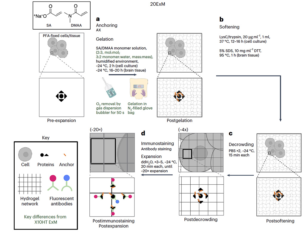
Expansion microscopy (ExM) is in increasingly widespread use throughout biology because its isotropic physical magnification enables nanoimaging on conventional microscopes. To date, ExM methods either expand specimens to a limited range (~4-10× linearly) or achieve larger expansion factors through iterating the expansion process a second time (~15-20× linearly). Here, we present an ExM protocol that achieves ~20× expansion (yielding <20-nm resolution on a conventional microscope) in a single expansion step, achieving the performance of iterative expansion with the simplicity of a single-shot protocol. This protocol, which we call 20ExM, supports postexpansion staining for brain tissue, which can facilitate biomolecular labeling. 20ExM may find utility in many areas of biological investigation requiring high-resolution imaging.