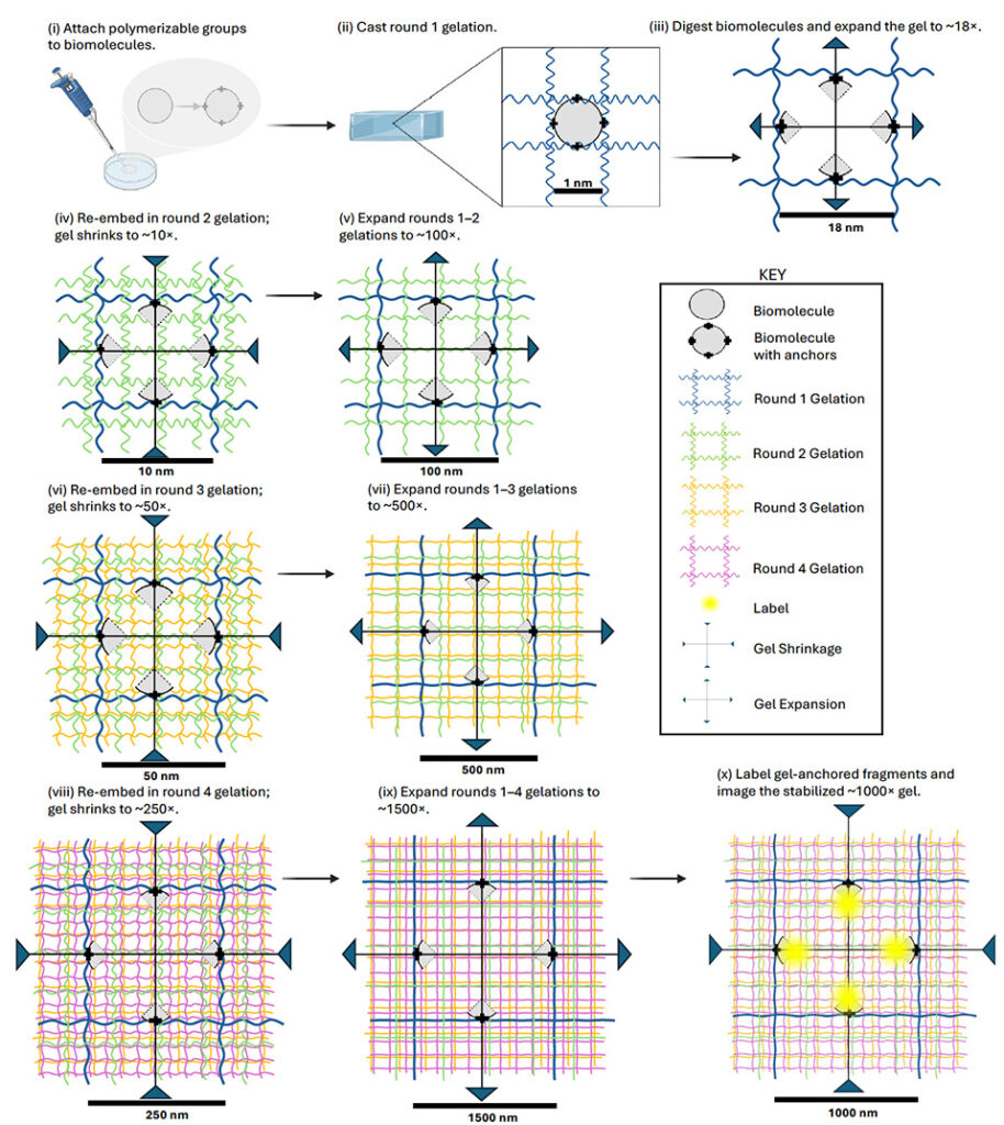
Biological macromolecules, such as proteins, are made of concatenated building blocks. We hypothesized that individual protein residues could be imaged by anchoring their side chains to a swellable polymer, cleaving backbone amide bonds, and expanding residues away from each other to a degree that enables them to be visualized separately. We introduce thousandfold expansion microscopy (1000ExM), a four-network interpenetrating hydrogel architecture that enables successive expansion from ∼18-fold to >1000-fold (one billion-fold in volume). Protein and peptide structures are maintained across these expansion factors, as verified by analyses of proteins with known structures (nanobodies, GFP) and a well-studied peptide (mCLING). Computational analysis indicates that 1000ExM resolves adjacent amino acid residues, thereby achieving sub-nanometer precision on conventional light microscopes. We anticipate that 1000ExM will find wide utility in protein visualization and identification, potentially even in intact cells and tissues.