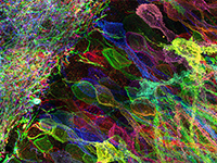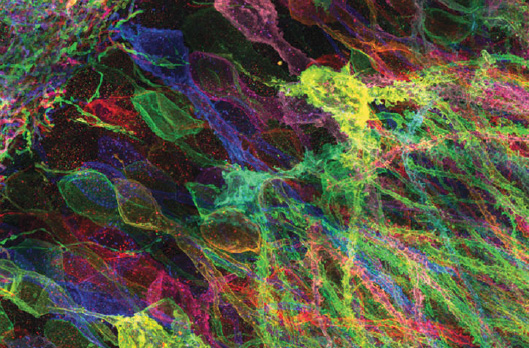
Expansion microscopy (ExM) is a recently invented technology that uses swellable charged polymers, synthesized densely and with appropriate topology throughout a preserved biological specimen, to physically magnify the specimen 100-fold in volume, or more, in an isotropic fashion. ExM enables nanoscale resolution imaging of preserved samples on inexpensive, fast, conventional microscopes. How does ExM work? How good is its performance? How do you get going on using it? In this Q&A, we provide the answers to these and other questions about this new and rapidly spreading toolbox.