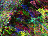
Hydrogels are extensively used as tunable, biomimetic three-dimensional cell culture matrices, but optically deep, high-resolution images are often difficult to obtain, limiting nanoscale quantification of cell-matrix interactions and outside-in signalling. Here we present photopolymerized hydrogels for expansion microscopy that enable optical clearance and tunable ×4.6-6.7 homogeneous expansion of not only monolayer cell cultures and tissue sections, but cells embedded within hydrogels. The photopolymerized hydrogels for expansion microscopy formulation relies on a rapid photoinitiated thiol/acrylate mixed-mode polymerization that is not inhibited by oxygen and decouples monomer diffusion from polymerization, which is particularly beneficial when expanding cells embedded within hydrogels. Using this technology, we visualize human mesenchymal stem cells and their interactions with nascently deposited proteins at <120 nm resolution when cultured in proteolytically degradable synthetic polyethylene glycol hydrogels. Results support the notion that focal adhesion maturation requires cellular fibronectin deposition; nuclear deformation precedes cellular spreading; and human mesenchymal stem cells display cell-surface metalloproteinases for matrix remodelling.
