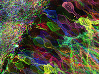
Nanoscale imaging of whole vertebrates is essential for the systematic understanding of human diseases, yet this goal has not yet been achieved. Expansion microscopy (ExM) is an attractive option for accomplishing this aim; however, the expansion of even mouse embryos at mid- and late-developmental stages, which have fewer calcified body parts than adult mice, is yet to be demonstrated due to the challenges of expanding calcified tissues. Here, we introduce a state-of-the-art ExM technique, termed whole-body ExM, that utilizes cyclic digestion. This technique allows for the super-resolution, volumetric imaging of anatomical structures, proteins, and endogenous fluorescent proteins (FPs) within embryonic and neonatal mice by expanding them 4-fold. The key feature of whole-body ExM is the alternating application of two enzyme compositions repeated multiple times. Through the simple repetition of this digestion process with an increasing number of cycles, mouse embryos of various stages up to E18.5, and even neonatal mice, which display a dramatic difference in the content of calcified tissues compared to embryos, are expanded without further laborious optimization. Furthermore, the whole-body ExM’s ability to retain FP signals allows the visualization of various neuronal structures in transgenic mice. Whole-body ExM could facilitate studies of molecular changes in various vertebrates.
