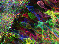
Determining the structure and composition of macromolecular assemblies is a major challenge in biology. Here we describe ultrastructure expansion microscopy (U-ExM), an extension of expansion microscopy that allows the visualization of preserved ultrastructures by optical microscopy. This method allows for near-native expansion of diverse structures in vitro and in cells; when combined with super-resolution microscopy, it unveiled details of ultrastructural organization, such as centriolar chirality, that could otherwise be observed only by electron microscopy.
