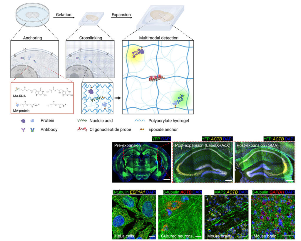
Expansion microscopy (ExM), by physically enlarging specimens in an isotropic fashion, enables nanoimaging on standard light microscopes. Key to existing ExM protocols is the equipping of different kinds of molecules, with different kinds of anchoring moieties, so they can all be pulled apart from each other by polymer swelling. Here we present a multifunctional anchor, an acrylate epoxide, that enables proteins and RNAs to be equipped with anchors in a single experimental step. This reagent simplifies ExM protocols and reduces cost (by 2-10-fold for a typical multiplexed ExM experiment) compared to previous strategies for equipping RNAs with anchors. We show that this united ExM (uniExM) protocol can be used to preserve and visualize RNA transcripts, proteins in biologically relevant ultrastructures, and sets of RNA transcripts in patient-derived xenograft (PDX) cancer tissues and may support the visualization of other kinds of biomolecular species as well. uniExM may find many uses in the simple, multimodal nanoscale analysis of cells and tissues.
Original biorxiv preprint: version 1 posted on June 19, 2022, and available [here]