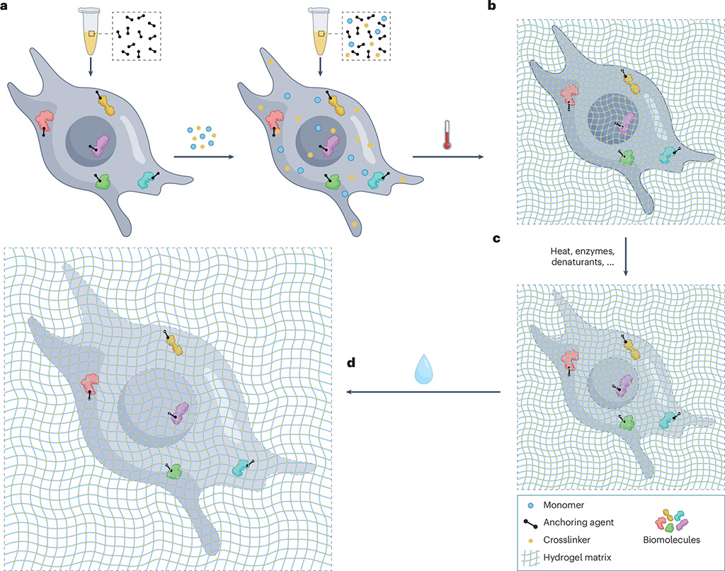
Expansion microscopy (ExM) is a sample transformation technique that enables the resolving of nanoscale biological features on ordinary microscopes. By physically expanding biological samples permeated by a hydrogel matrix, ExM effectively overcomes the diffraction limit of light microscopy, enabling nanoscale effective resolution on conventional imaging systems (such as confocal microscopes) without specialized super-resolution optics. Since its inception, ExM has evolved beyond fluorescence microscopy, enhancing the resolution of techniques such as mass spectrometry imaging, Raman imaging and in situ sequencing, by physically decrowding biomolecules. This Primer aims to provide a comprehensive guide to ExM, focusing on both its most common and latest variants. We outline a modular approach for understanding the scope of ExM protocols, as well as for understanding how to optimize or tune ExM for specific biological questions, covering essential laboratory equipment, sample preparation techniques, imaging techniques and computational approaches. Additionally, we discuss strategies for handling and analysing the vast data generated by ExM, highlight key applications across various biological fields, including cellular and structural biology, neuroscience, pathology and microbiology, and address current limitations. Finally, we explore emerging innovations in expansion chemistry, multimodal imaging and automation that will further broaden the impact of ExM across biological and clinical research.