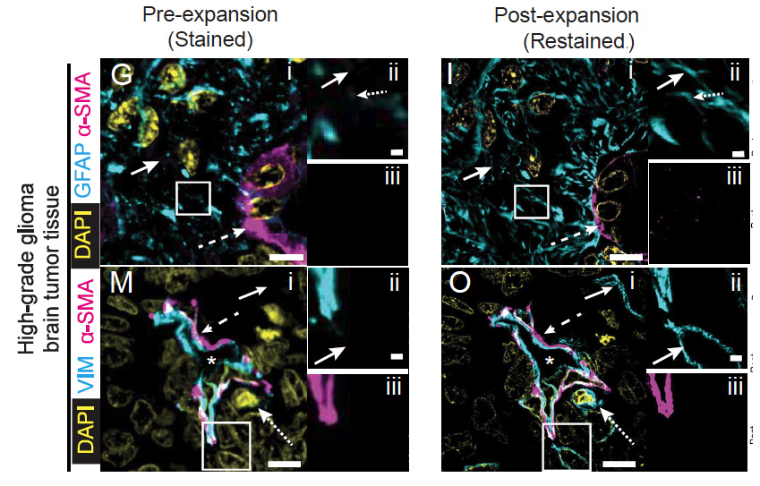
Proteins are densely packed in cells and tissues, where they form complex nanostructures. Expansion microscopy (ExM) variants have been used to separate proteins from each other in preserved biospecimens, improving antibody access to epitopes. Here we present an ExM variant, decrowding expansion pathology (dExPath), which can expand proteins away from each other in human brain pathology specimens, including formalin-fixed paraffin-embedded (FFPE) clinical specimens. Immunostaining of dExPath-expanded specimens reveals, with nanoscale precision, previously unobserved cellular structures, as well as more continuous patterns of staining. This enhanced molecular staining results in observation of previously invisible disease marker-positive cell populations in human glioma specimens, with potential implications for tumor aggressiveness. dExPath results in improved fluorescence signals even as it eliminates lipofuscin-associated autofluorescence. Thus, this form of expansion-mediated protein decrowding may, through improved epitope access for antibodies, render immunohistochemistry more powerful in clinical science and diagnosis.