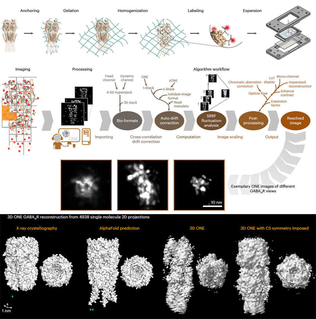
The attainable resolution of fluorescence microscopy has reached the subnanometer range, but this technique still fails to image the morphology of single proteins or small molecular complexes. Here, we expand the specimens at least tenfold, label them with conventional fluorophores and image them with conventional light microscopes, acquiring videos in which we analyze fluorescence fluctuations. One-step nanoscale expansion (ONE) microscopy enables the visualization of the shapes of individual membrane and soluble proteins, achieving around 1-nm resolution. We show that conformational changes are readily observable, such as those undergone by the ~17-kDa protein calmodulin upon Ca2+ binding. ONE is also applied to clinical samples, analyzing the morphology of protein aggregates in cerebrospinal fluid from persons with Parkinson disease, potentially aiding disease diagnosis. This technology bridges the gap between high-resolution structural biology techniques and light microscopy, providing new avenues for discoveries in biology and medicine.