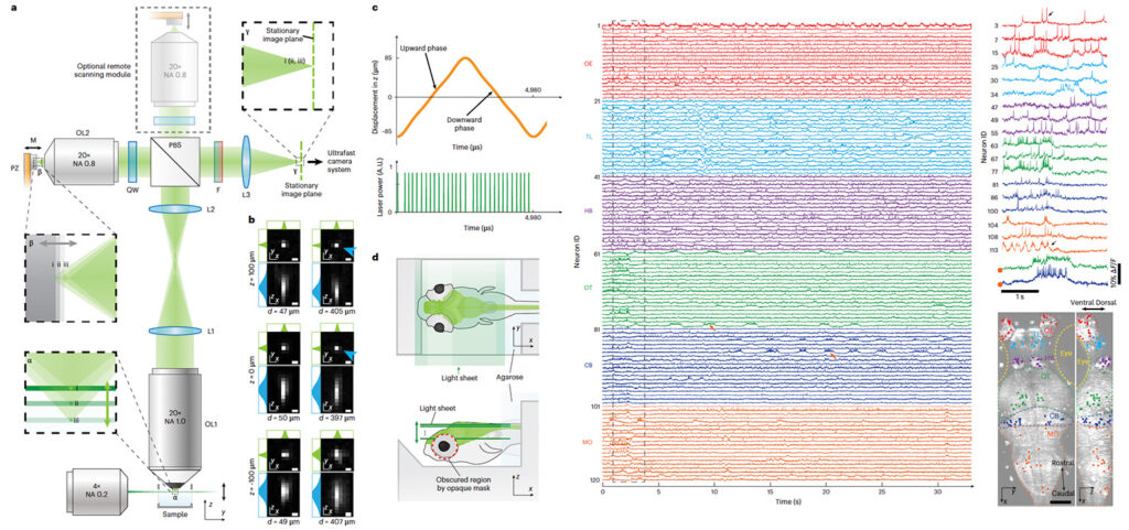
Neurons interact in networks distributed throughout the brain. While much effort has focused on whole-brain calcium imaging, advances in genetically encoded voltage indicators raise the question of whether it might be possible to image neuronal voltage across entire brains. Achieving this requires a microscope with high volumetric imaging rates and signal-to-noise ratio. Here we present a remote-scanning light-sheet microscope capable of imaging genetically encoded voltage indicator-expressing neurons distributed throughout much of the brain of larval zebrafish at a volumetric rate of 200.8 Hz. We measured voltage traces from approximately one-quarter of all brain neurons. We found that neurons firing at different times during a sequence occupied different locations: visually evoked sequences mapped across the optic tectum, whereas stimulus-independent bursts were mapped across the cerebellum and medulla. Imaging voltage of neurons distributed in many brain regions may open new frontiers for understanding fundamental neural system operations.
Original biorxiv preprint: version 1 posted on December 16, 2023, and available [here]