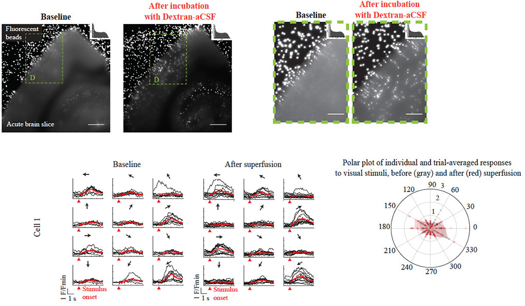
Established methods for imaging the living mammalian brain have, to date, taken the brain’s optical properties as fixed; we here demonstrate that it is possible to modify the optical properties of the brain itself to significantly enhance at-depth imaging while preserving native physiology. Using a small amount of any of several biocompatible materials to raise the refractive index of solutions superfusing the brain prior to imaging, we could increase severalfold the signals from the deepest cells normally visible and, under both one-photon and twophoton imaging, visualize cells previously too dim to see. The enhancement was observed for both anatomical and functional fluorescent reporters across a broad range of emission wavelengths. Importantly, visual tuning properties of cortical neurons in awake mice, and electrophysiological properties of neurons assessed ex vivo, were not altered by this procedure.
Original biorxiv preprint: version 1 posted on September 7, 2024, and available [here]