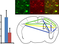
Dissecting neural circuitry in non-human primates (NHP) is crucial to identify potential neuromodulation anatomical targets for the treatment of pharmacoresistant neuropsychiatric diseases by electrical neuromodulation. How targets of deep brain stimulation (DBS) and cortical targets of transcranial magnetic stimulation (TMS) compare and might complement one another is an important question. Combining optogenetics and tractography may enable anatomo-functional characterization of large brain cortico-subcortical neural pathways. For the proof-of-concept this approach was used in the NHP brain to characterize the motor cortico-subthalamic pathway (m_CSP) which might be involved in DBS action mechanism in Parkinson’s disease (PD). Rabies-G-pseudotyped and Rabies-G-VSVg-pseudotyped EIAV lentiviral vectors encoding the opsin ChR2 gene were stereotaxically injected into the subthalamic nucleus (STN) and were retrogradely transported to the layer of the motor cortex projecting to STN. A precise anatomical mapping of this pathway was then performed using histology-guided high angular resolution MRI tractography guiding accurately cortical photostimulation of m_CSP origins. Photoexcitation of m_CSP axon terminals or m_CSP cortical origins modified the spikes distribution for photosensitive STN neurons firing rate in non-equivalent ways. Optogenetic tractography might help design preclinical neuromodulation studies in NHP models of neuropsychiatric disease choosing the most appropriate target for the tested hypothesis.
