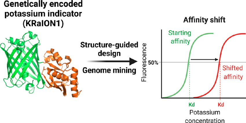
Genetically encoded potassium indicators lack optimal binding affinity for monitoring intracellular dynamics in mammalian cells. Through structure-guided design and genome mining of potassium binding proteins, we developed green fluorescent potassium indicators with a broad range of binding affinities. KRaION1 (K+ ratiometric indicator for optical imaging based on mNeonGreen 1), based on the insertion of a potassium binding protein, Kbp, from E. coli (Ec-Kbp) into the fluorescent protein mNeonGreen, exhibits an isotonically measured Kd of 69 ± 10 mM (mean ± standard deviation used throughout). We identified Ec-Kbp’s binding site using NMR spectroscopy to detect protein–thallium scalar couplings and refined the structure of Ec-Kbp in its potassium-bound state. Guided by this structure, we modified KRaION1, yielding KRaION1/D9N and KRaION2, which exhibit isotonically measured Kd’s of 138 ± 21 and 96 ± 9 mM. We identified four Ec-Kbp homologues as potassium binding proteins, which yielded indicators with isotonically measured binding affinities in the 39–112 mM range. KRaIONs functioned in HeLa cells, but the Kd values differed from the isotonically measured case. We found that, by tuning the experimental conditions, Kd values could be obtained that were consistent in vitro and in vivo. We thus recommend characterizing potassium indicator Kd in the physiological context of interest before application.
Original biorxiv preprint: version 1 posted on October 8, 2021, and available [here]
